Scope
Only 12.3 A–C in the supplied document. Sections 12.1, 12.2 and later 12.3 sections are excluded. Existing source tabs and pictures are preserved.
Only A–C. Complete explanations, key points and shared visual posters.
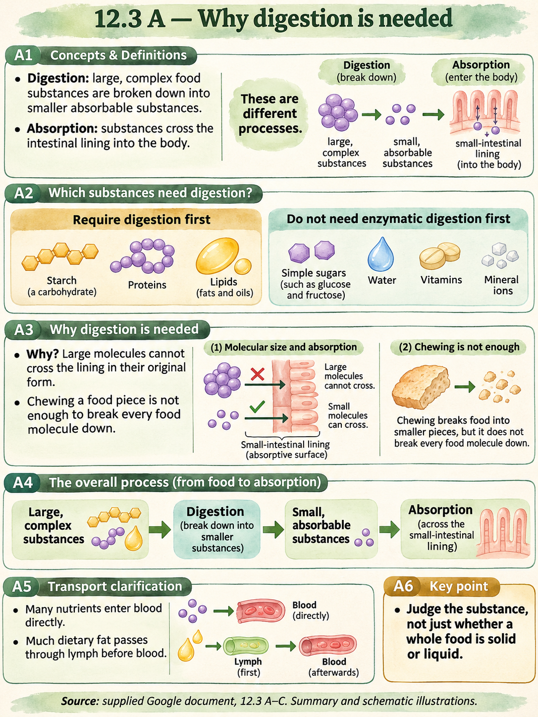
Digestion breaks large, complex food molecules into smaller, simpler substances. Absorption is the movement of absorbable substances across the intestinal wall into the body. These are different processes.
Starch, proteins and lipids require digestion before their products can be absorbed. Simple sugars such as glucose and fructose, water, vitamins and mineral ions do not need enzymatic digestion first.
Large food molecules cannot pass through the intestinal lining in their original form. Digestion produces smaller substances that can cross it. Crushing a food piece alone does not necessarily make its individual molecules small enough.
The source uses a simplified blood-transport account. Many absorbed nutrients enter blood directly; much dietary fat is transported via lymph before reaching blood. For A–C, the main principle is digestion before absorption for large food substances, not the detailed transport pathways.
Minerals are described more precisely as mineral ions rather than calling every listed substance a simple molecule. No digestion is needed for water or mineral ions.
| Food substance | Digestion needed first? | Reason |
|---|---|---|
| Starch, proteins, lipids | Yes | Their original large forms must be broken down. |
| Simple sugars | No | Already small enough for absorption. |
| Water, vitamins, mineral ions | No enzymatic digestion first | They can be absorbed without being broken down by digestive enzymes. |
The digestive system is a group of organs working together to make food substances available for absorption. It consists of the alimentary canal and associated glands.
The alimentary canal is the continuous tube through which food travels. Glands produce secretions that help digestion; food does not travel through each gland.
A complex function requires coordinated organs: food must be moved and physically broken up, digestive secretions must reach it, and useful products must become available for absorption.
A large piece of food and a large molecule are not the same thing. Mechanical actions reduce piece size; chemical digestion changes molecules. Both contribute to the overall function.
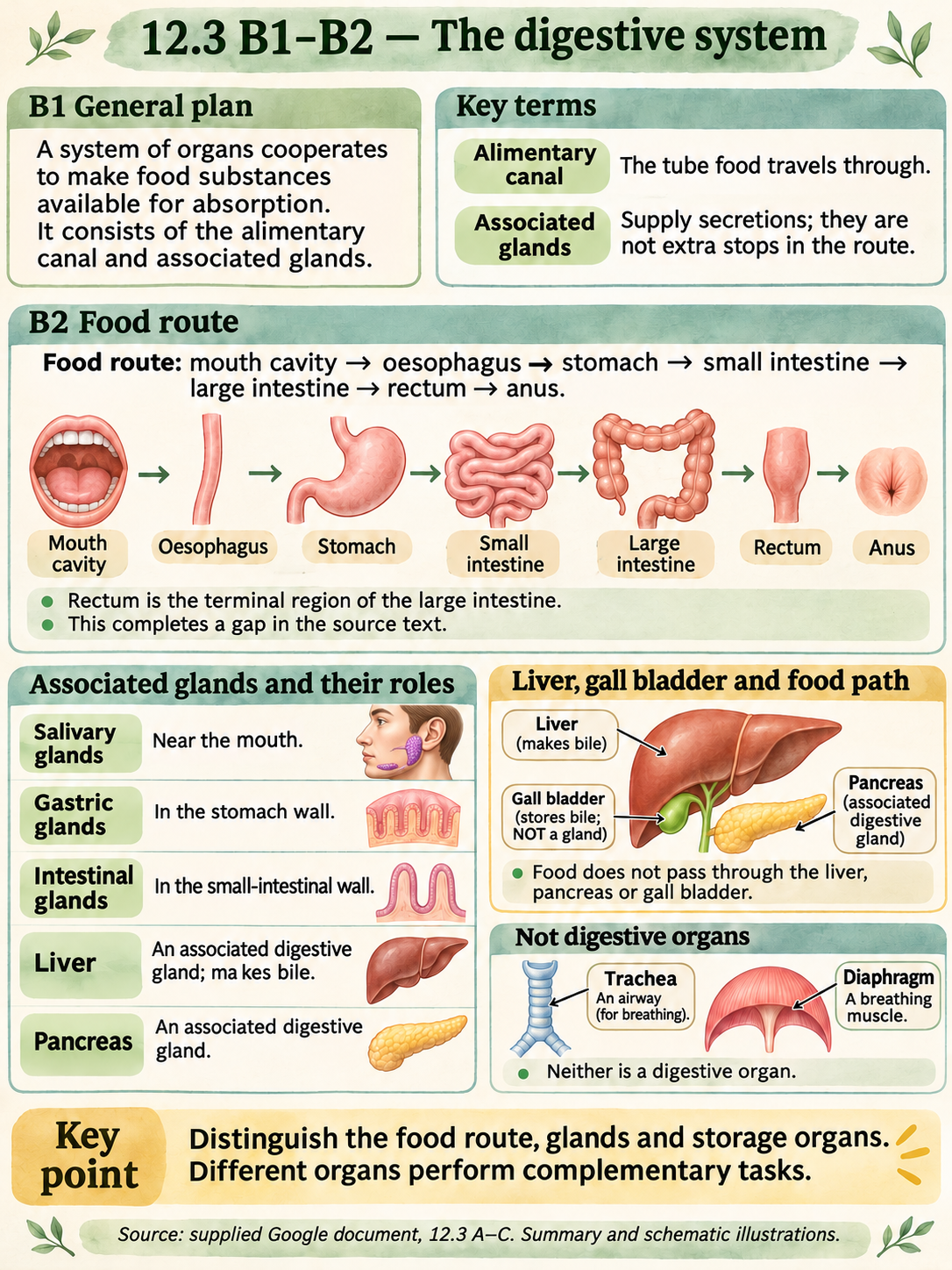
Food route: mouth cavity → oesophagus → stomach → small intestine → large intestine → rectum → anus. The rectum is the terminal storage region of the large intestine.
Associated glands include salivary glands, gastric glands in the stomach wall, the liver, the pancreas, and intestinal glands in the small-intestinal wall. The gall bladder stores bile made by the liver; it is not itself a gland.
Food stays within the alimentary canal. Secretions reach food from associated organs; the liver, pancreas and gall bladder are not additional stops in the food route.
The trachea is an airway and the diaphragm is a breathing muscle. They may appear near the digestive organs in a diagram but are not parts of the digestive system.
The source text leaves the route blank. The route has been reconstructed from its labelled digestive-system picture and checked against the NIDDK overview; the rectum is explicitly identified as a clarification.
| Structure | Category or role |
|---|---|
| Mouth, oesophagus, stomach, intestines, rectum, anus | Parts or regions of the alimentary canal |
| Salivary, gastric and intestinal glands; liver; pancreas | Associated digestive glands |
| Gall bladder | Stores bile from the liver; not a gland |
| Trachea; diaphragm | Not part of the digestive system |
Mechanical digestion physically reduces food-piece size. The food substance is not chemically changed by that physical action alone.
Chemical digestion uses digestive enzymes to speed up reactions that break complex food molecules into simpler products. The chemical structure changes. Different enzymes act on different food substances.
Breaking a food piece into smaller pieces increases the total surface area exposed to digestive juice. More contact helps enzymes act on the food; chewing does not itself turn starch into sugar.
In the source summary, starch is broken down to sugars; proteins are broken down through smaller chains such as polypeptides to amino acids; lipids yield fatty acids and glycerol in the simplified school model.
A digestive secretion is not automatically an enzyme. For example, bile is a secretion made by the liver, so the statement that every digestive juice contains enzymes is too broad.
| Feature | Mechanical digestion | Chemical digestion |
|---|---|---|
| Main change | Food pieces become smaller | Complex molecules are broken down |
| Chemical structure | Unchanged by the physical action itself | Changed |
| Main benefit | Greater contact with digestive juice | Produces smaller, absorbable substances |
| Mouth example | Chewing with teeth | Salivary amylase acting on starch |
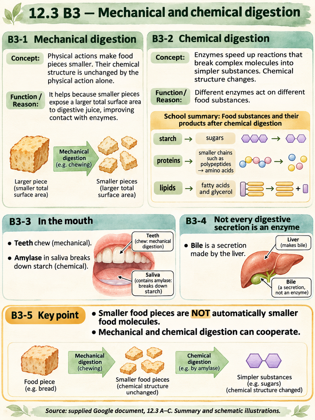
Both mechanical and chemical digestion occur in the mouth. Teeth chew food into smaller pieces. Salivary glands produce saliva containing the enzyme amylase.
Amylase breaks starch into simpler sugars. Chewing and amylase action are different processes even when they happen at the same time.
Chewing exposes a larger total food surface to saliva, helping contact between food and digestive juice. It changes piece size, not the starch molecule by itself.
Amylase speeds up starch breakdown. The presence of amylase does not mean that all food substances are digested by that same enzyme.
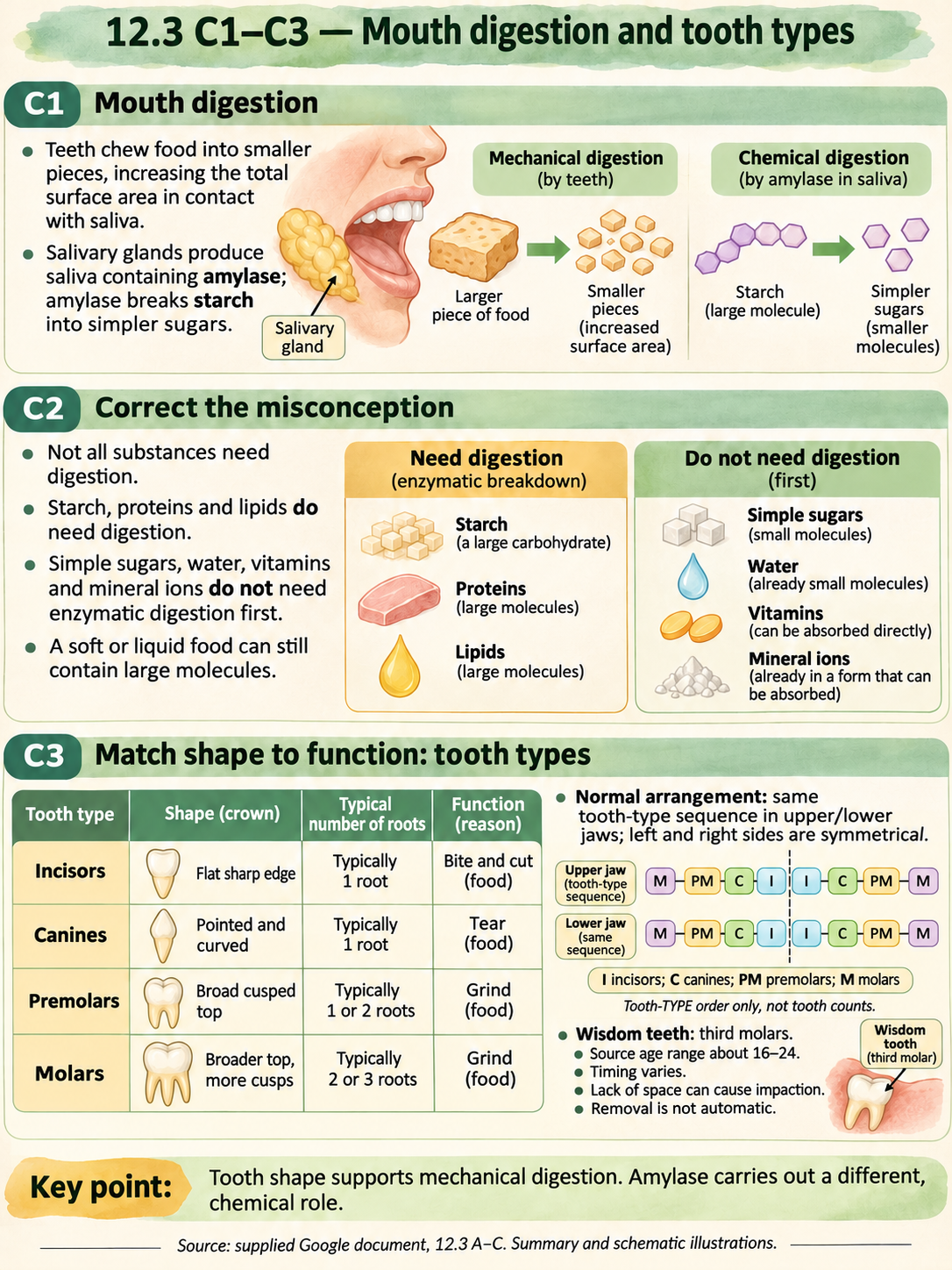
The claim that every food substance must be digested before absorption is incorrect. The need depends on the food substance, not simply on whether a whole food has been swallowed.
One food or drink can contain both substances needing digestion and substances that do not. For example, starch and water in the same food have different requirements.
Starch, proteins and lipids have to be broken down. Simple sugars, vitamins, water and mineral ions can be absorbed without enzymatic digestion first.
A food being soft, crushed, liquid or dissolved does not establish that all of its molecules are small enough for absorption. Piece size and molecular size must be considered separately.
The four tooth types are incisors, canines, premolars and molars. Their shapes suit different mechanical actions.
The source gives a symmetrical left/right arrangement, with the same tooth-type sequence in the upper and lower jaws. This describes the normal plan, not identical shape of every tooth.
Wisdom teeth are third molars. The source gives about ages 16–24 for their appearance. Timing and presence vary. Lack of space can cause sideways or impacted growth; removal is a clinical decision, not inevitable.
A flat sharp incisor edge helps bite and cut. A pointed canine helps tear. Broad cusped premolar and molar surfaces help crush and grind, increasing exposed food surface.
Root counts in the source are typical teaching patterns, not a rule that every tooth has the same root anatomy. They help describe structure but shape and position must also be considered.
| Type | Typical shape | Source root pattern | Function |
|---|---|---|---|
| Incisor | Flat sharp edge | 1 | Biting and cutting |
| Canine | Pointed, curved | 1 | Tearing |
| Premolar | Broad top with cusps | 1 or 2 | Grinding |
| Molar | Broader top, more cusps | 2 or 3 | Grinding |
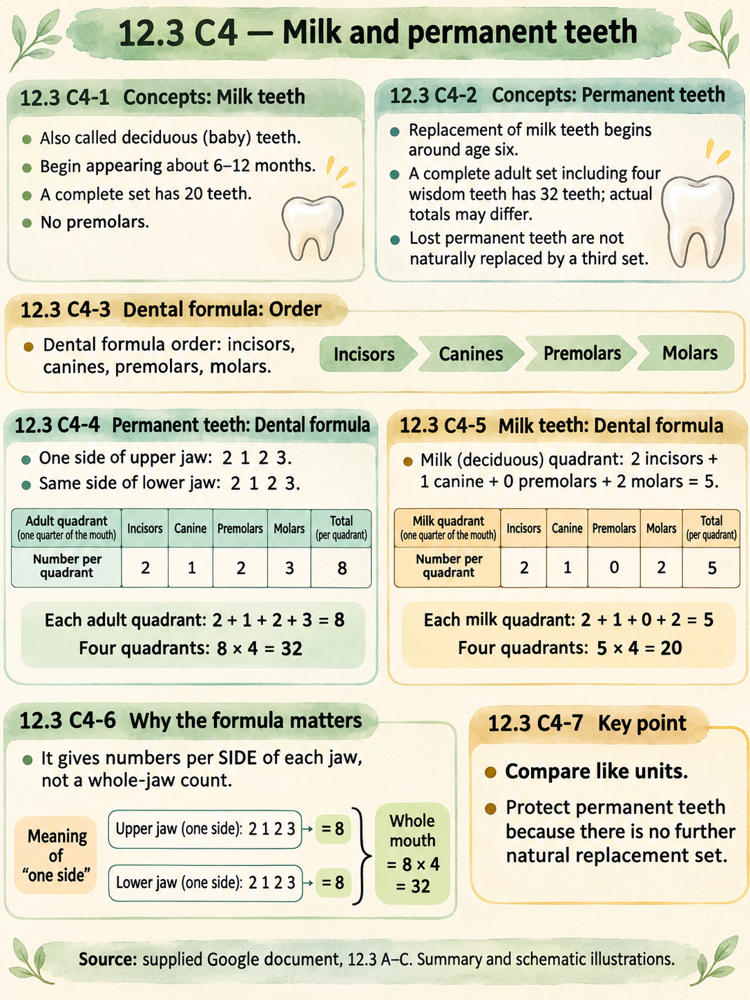
Milk teeth begin appearing at about 6–12 months in the source account. A complete milk set has 20 teeth and no premolars.
Permanent teeth begin replacing milk teeth around age six. A complete adult arrangement including four wisdom teeth has 32 teeth; actual numbers can differ if wisdom teeth are absent or removed.
A dental formula shows tooth numbers on one side of the upper jaw above those on the same side of the lower jaw. The adult order is incisors, canines, premolars, molars.
The adult formula is 2,1,2,3 over 2,1,2,3. One quadrant has 2+1+2+3=8 teeth; four quadrants give 32. It is not a formula for the entire jaw.
The milk arrangement has 2 incisors, 1 canine, 0 premolars and 2 molars per quadrant: 2+1+0+2=5; 5×4=20. This is a calculated explanation of the source description.
A lost permanent tooth is not naturally replaced by another normal set. Protecting permanent teeth matters because there is no third replacement dentition.
| Per quadrant | Incisors | Canines | Premolars | Molars | Total |
|---|---|---|---|---|---|
| Milk | 2 | 1 | 0 | 2 | 5 |
| Permanent, including wisdom tooth | 2 | 1 | 2 | 3 | 8 |
Plaque is a sticky bacterial film on teeth. Tooth decay and periodontal disease both involve plaque bacteria but damage different structures through different processes.
Tooth decay begins when acids produced as plaque bacteria act on food debris damage the hard mineralised enamel. Decay may extend through dentine into the pulp cavity, which contains nerves and blood vessels.
Periodontal disease affects the gums and supporting tissues. Gum inflammation can cause swelling and separation from teeth, forming pockets. Progression may damage cementum, periodontal membrane and jawbone.
Acid dissolves enamel mineral, allowing a cavity to form. If decay reaches the pulp, irritation of nerves can cause pain; infection may cause pus at the root tip. Pain is not a necessary early sign.
Loss of supporting tissues in periodontal disease reduces the stability of teeth, so they may loosen or be lost. This differs from describing all oral disease as simply an enamel hole.
| Feature | Tooth decay | Periodontal disease |
|---|---|---|
| Early target | Enamel / tooth substance | Gums and supporting tissues |
| Main chain | Bacterial acid → mineral loss → cavity | Plaque → inflammation → support damage |
| Advanced consequence | Pulp involvement and infection | Loosening or loss of teeth |
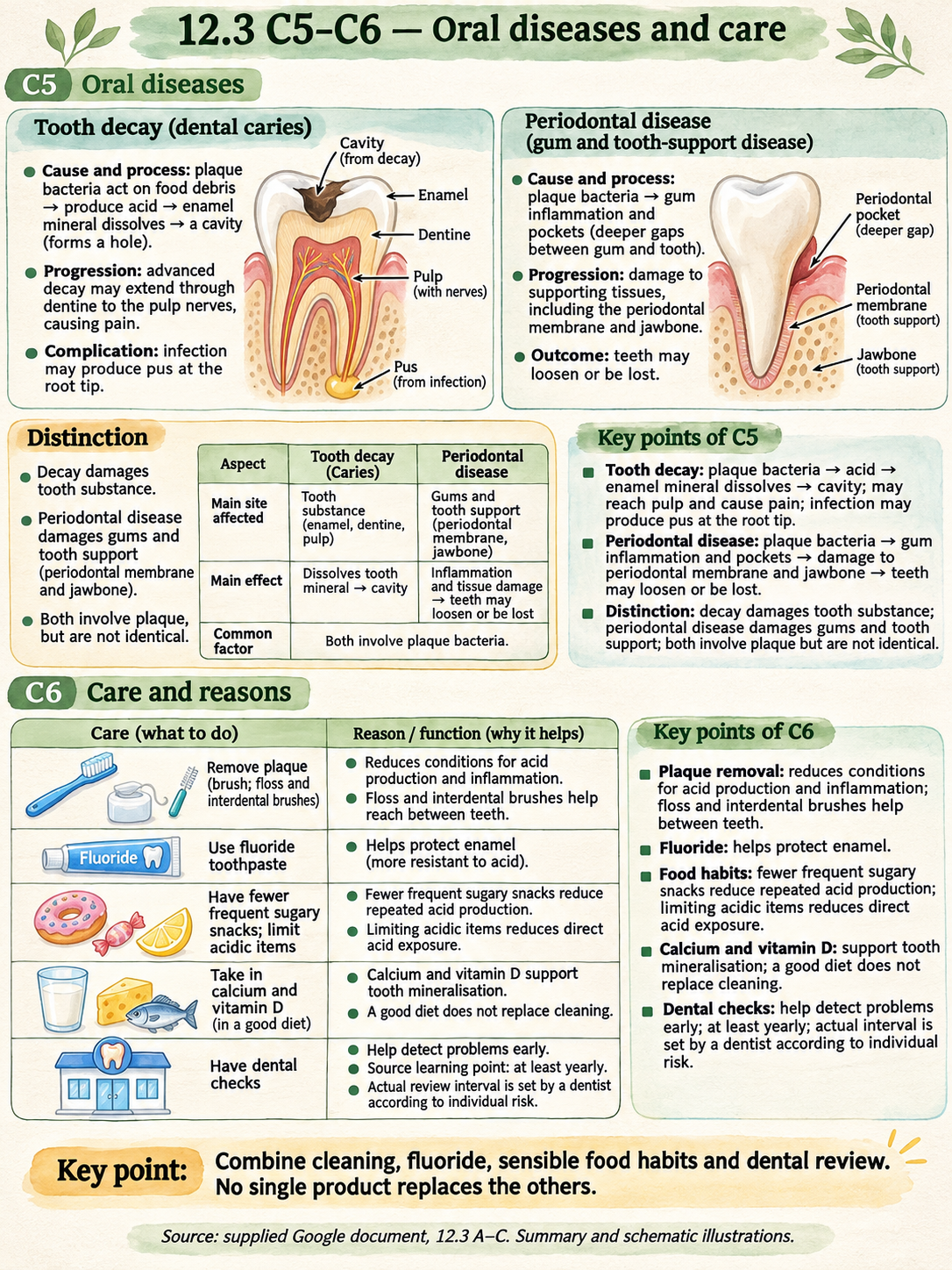
The source lists dental check-ups, calcium- and vitamin-D-rich foods, reduced sugary/acidic foods and eating frequency, fluoride toothpaste, and appropriate interdental cleaning products.
These measures complement one another. Cleaning the tooth surface, cleaning between teeth and reducing repeated sugar or acid exposure address different parts of the disease process.
Removing plaque and trapped debris reduces conditions that support acid production and inflammation. Interdental brushes and floss reach between teeth where an ordinary brush may miss areas.
Fluoride helps protect enamel against decay. Reducing frequent sugary snacks reduces repeated opportunities for bacterial acid production; limiting acidic items reduces direct acid exposure.
Calcium and vitamin D support tooth mineralisation. Diet does not replace plaque removal. Dental checks help identify problems early.
The source says at least one check-up per year. This is retained as a source learning point, not a universal clinical interval: actual recall is set by a dentist according to individual oral health and risk.
Only 12.3 A–C in the supplied document. Sections 12.1, 12.2 and later 12.3 sections are excluded. Existing source tabs and pictures are preserved.
The supplied document contains summaries and previously generated pictures rather than a complete paginated textbook scan. Source locations use tab names; printed page numbers are not invented.
The source text has a blank route after its route heading. The source labelled picture supplies mouth, oesophagus, stomach, small and large intestines, anus. The route is reconstructed; rectum is added explicitly as the terminal large-intestinal region, checked against NIDDK.
The source says digestion products enter blood directly. This is a simplified account; much absorbed fat reaches blood through lymph first. The learning note avoids saying every nutrient goes directly into blood.
Minerals are called mineral ions, rather than describing every listed substance as a molecule. This is a scientific wording clarification.
The source generalises that digestive juices contain enzymes. This is qualified: enzyme-containing secretions carry out chemical digestion; bile is a secretion but not an enzyme.
One B mechanical/chemical picture shows a pancreas-like drawing labelled salivary gland, and a general-plan picture can suggest absorption from the stomach. New diagrams distinguish glands correctly and show the intestinal lining for the A absorption principle.
Removal is phrased as may be needed after dental assessment, not inevitable. Source age and root patterns are presented as typical textbook ranges.
Adult 2.1.2.3 per quadrant and complete total 32 are read from the source English PIC. Milk 2.1.0.2 and 20 are explained by calculation, not claimed as a newly recovered quotation.
Source annual-check language is distinguished from an individual recommendation; NHS describes risk-based intervals.
All exercise scenarios, model observations and datasets are newly authored educational examples. They are not personal diagnoses or actual class experimental results.